Temporomandibular Joint Disorders
Temporomandibular Joint Disorders

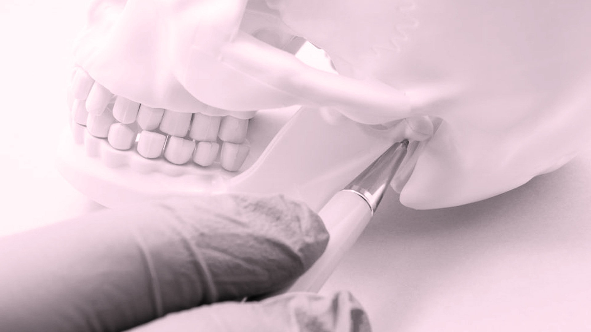
The temporomandibular joint (TMJ) is the articulation of the lower jaw to the temporal bone skull. It acts as a sliding hinge and controls the movement of the jaw down, up, and to the side. There are two joints one on each side located in front of the ears. TMJs are parts of the masticatory system which also includes the teeth, the jaws and the muscles which control the movement of the lower jaw. Any condition affecting a part of the masticatory system may also influence the function of the other parts. So the masticatory system must be evaluated as a whole in order to evaluate and better understand its dysfunction.
In General temporomandibular joint disorders are described as pain of the muscles or of the joint itself. The exact cause of a patient’s TMJ disorder is often difficult to determine. Multiple factors may be considered as possible causes such as jaw injury, genetics, arthritis, or clenching or grinding of the teeth, changes in occlusion due to dental treatments, missing teeth, stress.
Symptoms of Tmj disorder usually include pain or tenderness of the jaw, pain in one or both of the temporomandibular joints, pain in and around your ear, difficulty in chewing or biting, facial pain radiating to the head or to the neck, limitation of the joint movement making it difficult to open or close the mouth (locking), earaches and ringing in the ears (tinnitus), clicking, popping or a grating sound in the temporomandibular joints(crepitus).
The mandibular condyle within the temporomandibular joint (TMJ) can rotate (hinge action) and slide forward. The bony parts that interact as the joint functions constantly are covered with cartilage and separated by a shock-absorbing disk, which normally follows the movements of the condyle. The most frequent cause of TMJ dysfunction is internal derangement which refers to an alteration in the normal pathways of motion of the articular disc. TMJ disorders can occur if: The disk erodes or moves out of its proper alignment (usually it moves forward and medial before the movement of the condyle), arthritis damages the joint's cartilage, or an impact damages the joint.
Pain or discomfort involving the temporomandibular joints is not uncommon. Studies have shown that 10-30% of the general population may exhibit symptoms associated with TMJ disorders. Many times patients will seek treatment from other specialists such as ENTs or Neurologists as the symptoms may mimic ear problems, migraines or neuralgia.
The diagnosis is usually made by the combination of patient’s history, clinical exam and radiographic evaluation. The description of the condition from various patients is many times identical, although there may be different clinical scenarios. The clinician will evaluate the range of jaw motion, the condition of the dental arches, the tenderness on palpation of the masticatory muscles, the noises heard during the joint function, the tenderness of the joint while pressing on it. Other helpful diagnostic modalities may include simple x-rays to evaluate whether the bony structures of the joints are intact, CT scans for a more detailed look of the bone surfaces and MRIs to evaluate the condition as well as the position of the articular disc. In few cases, direct visualization of the joint with arthroscopy may be necessary.
Quite often, the discomfort associated with TMJ disorders is temporary and can be relieved with simple self-managed measures or conservative treatments. Surgery is typically reserved for cases that conservative management has failed.
Simple home measures include: Anti-inflammatory over the counter medications or analgesics, application of heat packs, avoidance of excessive jaw movement, and soft food diet.
Conservative treatments include: Prescribed anti-inflammatory medications or analgesics, occlusal splints provided by prosthodontists or specialized dentists, physiotherapy of the facial muscles, correction of any occlusal or dental problems, trigger point injection of analgesics, Botox injections to the muscles of mastication.
Surgical treatment is undertaken under certain criteria. The patient has followed a conservative treatment path with no signs of improvement. The CT reveals significant bony changes due to chronic overload and dysfunction of the joints, osteoarthritic alterations or resorption of the condyle. The MRI shows disk displacement or disk morphology and shape alterations. Even in the presence of the above findings, the decision for surgery will be based on the patient’s quality of life as well as the level of function. Surgery will not usually be a first line treatment if the patient has minimal or moderate symptoms.
Surgical procedures that we provide in our clinic for the treatment of TMJ disorders include:
Arthrocentesis. Under local anesthesia or sedation, two catheters are inserted into the joint space. Fluid is irrigated through the joint to remove any inflammatory by-products and joint debris. At the same time, the surgeon moves the jaw in multiple positions aiming for lysis of adhesions within the joint and to facilitate the positioning of the displaced disk into its normal position. It is a minimally invasive procedure commonly used for cases of internal derangement or closed locks with very good results. The patient is discharged the same day.
The modified condylotomy is a surgical procedure used to manage patients with temporomandibular joint dysfunction. The purpose of the procedure is to increase joint space by allowing the mandibular condyle to move inferiorly.
Arthroplasty is a general term that refers to various surgical procedures of the Temporomandibular Joint. They are indicated for patients with progressively debilitating internal derangement refractory to the non-surgical and minimally invasive techniques described above. Other indications include degenerative joint disease, the treatment of various pathologic processes and ankylosis.
Condylectomy refers to the removal of the condylar head. It is an open joint procedure.
High condylectomy (condylar shave) with and without replacement is a procedure during which we remove the upper usually eroded part of the condylar head.
Disk plication with Mitec Anchors. This is a surgical procedure during which the anteriorly displaced articular disk is pulled back into position as sutured to the condyle with the aid of a small anchor which is placed on the head of the condyle.
Discectomy/meniscectomy and disc replacement. During this procedure, we remove the severely damaged articular disk. To prevent bone to bone contact and prevent TMJ ankylosis we fill the resultant gap with autogenous regional tissue graft from the temporalis area.
Eminectomy (removal of the articular eminence) is a surgical procedure used to eliminate the blocking factor preventing the mandibular condyle from returning to its rest position within the glenoid fossa. It is mainly used for the treatment of chronic dislocation of the condyle.
Total joint reconstruction. In patients with severe osteoarthritis, inflammatory arthrosis, connective or autoimmune disease, ankylosis, absent or deformed structures, congenital deformities, and sometimes chronic pain we may choose to perform a total joint reconstruction. In our clinic, the preferred method is to use a custom-made prosthetic joint. A stereolithographic model of the patient's skull is obtained, and the planned procedure is first done on this model. Then the model is sent to the laboratory, and a custom-made titanium polyethylene prosthesis is fabricated. This prosthesis will be used in the operating room to replace the diseased joint. It is a highly demanding technically sensitive procedure that needs training and experience from the surgical team.
The most common reason for an unsuccessful TMJ surgery is a failure to eliminate the underlying cause of the problem in addition to the obvious pathology. When the surgeon deals only with the immediate situation and fails to consider what initially led to its development, there is a risk of the same cause will lead to a recurrence of the condition.
Another reason why TMJ surgery may not be successful is a wrong preoperative diagnosis. The signs and symptoms in patients with masticatory system dysfunction and those with other diseases of the TMJ may be analogous. The common etiologic relationship between muscle and joint problems may also lead to a mistaken assessment. Furthermore, even if a correct diagnosis is made, the patient may be treated with surgery although conservative management might have had a satisfactory outcome.
It is for these reasons that we insist on very careful evaluation and treatment planning for our patients with TMJ disorders. Although the arsenal of our surgical experience includes many surgical approaches for their treatment, we prefer to exhaust conservative treatment before we decide that the only option left is the surgical treatment.


